The visceral surface or inferior surface of the liver is concave, directed downward, backward, and to the left. Deep fissures and the impressions of the adjacent organs make this surface uneven. These two surfaces join antero-laterally in acute angle forming the inferior margin.
What are the surfaces of the liver?
Superior, anterior, posterior and right surfaces of the liver are continuous with each other and are related to the diaphragm and anterior abdominal wall. The anterior surface is separated from the inferior (visceral) surface by a sharp anterior (inferior) border that is clinically palpable on deep inspiration.
Which structure is not seen in visceral surface of the liver?
The surface is almost completely invested by peritoneum; the only parts devoid of this covering are where the gall-bladder is attached to the liver, and at the porta hepatis where the two layers of the lesser omentum are separated from each other by the bloodvessels and ducts of the liver.
What organs have a contact to the visceral surface of the liver?
Visceral Surface Relations Behind the colic impression is the renal impression, produced by the right kidney and right adrenal gland. Fat, connective tissue, and peritoneum intervene between these organs and the liver. The right adrenal gland is in contact with the bare area of the liver.What is the diaphragmatic surface of liver?
Diaphragmatic surface – the anterosuperior surface of the liver. It is smooth and convex, fitting snugly beneath the curvature of the diaphragm.
Where is your porta hepatis?
The porta hepatis, or hilum of the liver, is a deep, short, transverse fissure that passes across the left posterior aspect of the undersurface of the right lobe of the liver. It separates the caudate lobe and process from the quadrate lobe and meets the left sagittal fossa perpendicularly (1).
What holds the liver in place?
A layer of fibrous tissue called Glisson’s capsule covers the outside of the liver. This capsule is further covered by the peritoneum, a membrane that forms the lining of the abdominal cavity. This helps hold the liver in place and protects it from physical damage.
Is the liver intraperitoneal?
Intraperitoneal organs include the stomach, spleen, liver, first and fourth parts of the duodenum, jejunum, ileum, transverse, and sigmoid colon.Is the liver inferior to the gallbladder?
The gall bladder is adhered to the inferior surface of the liver. Its inferior surface is covered by a layer of the peritoneum. The quadrate lobe of the liver lies between the fissure for the ligamentum teres and the gallbladder [1].
What are lobules in the liver?Liver lobules are collections of hepatocytes in a hexagonal shape with the center being a central vein. … Blood flows from the portal vein branch and hepatic artery branch across the lobule and finally into the central vein which is a branch of the hepatic vein.
Article first time published onWhat is posterior to the liver?
The caudate lobe (lobus caudatus; Spigelian lobe) is situated upon the posterior surface of the right lobe of the liver, opposite the tenth and eleventh thoracic vertebræ. It is bounded, below, by the porta; on the right, by the fossa for the inferior vena cava; and, on the left, by the fossa for the ductus venosus.
What are the four lobes of the liver?
Anatomically the liver has four lobes: right, left, caudate, and quadrate. The quadrate lobe is located on the inferior surface of the right lobe. The caudate lobe is located between the left and right lobes in an anterior and superior location.
What is the name given to the region of the liver that is not covered by peritoneum?
Anatomical terminology The bare area of the liver (nonperitoneal area) is a large triangular area on the diaphragmatic surface of the liver, devoid of peritoneal covering. It is attached directly to the diaphragm by loose connective tissue.
What is visceral surface?
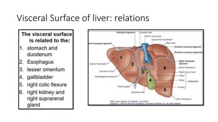
The visceral surface (inferior surface), is uneven, concave, directed downward, backward, and to the left, and is in relation with the stomach and duodenum, the right colic flexure, and the right kidney and suprarenal gland.
Is the liver attached to the diaphragm?
The liver is located under the ribs on the right hand side of the body. It lies just below the lungs, under the top of the diaphragm to which it is attached. The diaphragm is the muscle beneath the lungs which regulates our breathing. The liver is partly protected by the rib cage.
How is the liver attached?
The left and right lobe are divided by the falciform (“sickle-shaped” in Latin) ligament, which connects the liver to the abdominal wall. The liver’s lobes can be further divided into eight segments, which are made up of thousands of lobules (small lobes).
What are the 5 ligaments that anchor the liver?
The liver has several ligamentous attachments that anchor it in the right upper quadrant (RUQ) of the abdominal cavity. These include the coronary, triangular, falciform, round, hepatogastric and hepatoduodenal ligaments, together with ligamentum venosum.
Is the liver attached to anything?
The liver consists of 2 main lobes. Both are made up of 8 segments that consist of 1,000 lobules (small lobes). These lobules are connected to small ducts (tubes) that connect with larger ducts to form the common hepatic duct.
What do the hepatocytes do?
Hepatocytes, the major parenchymal cells in the liver, play pivotal roles in metabolism, detoxification, and protein synthesis.
What are the 4 main contents of the porta hepatis?
The porta hepatis is the central intraperitoneal fissure of the liver that separates the caudate and the quadrate lobes. It is the entrance and the exit for several important vessels including the portal vein, the hepatic artery, the hepatic nervous plexus, the hepatic ducts and the lymphatic vessels.
What are the hepatic ducts?
A tube that carries bile from the liver. It starts where the right and left hepatic (liver) ducts join outside the liver. It ends where the cystic duct from the gall bladder joins it to form the common bile duct.
What is meant by porta hepatis?
The porta hepatis is a deep fissure in the inferior surface of the liver through which all the neurovascular structures (except hepatic veins) and hepatic ducts enter or leave the liver 1. It runs in the hepatoduodenal ligament and contains: common hepatic ducts (anterior to the right)
Is the liver superior to the duodenum?
The first segment of the duodenum—the superior part of the duodenum (called the duodenal bulb) is connected to the liver via the hepatoduodenal ligament. This connection allows for transportation of nutrients from the small intestine to the liver; it also allows the duodenum to receive bile from the liver.
Is the liver anterior or posterior to the gallbladder?
The gallbladder is located inferior (below) and posterior (behind) to the liver in the upper right quadrant (section) of the abdomen. It lies in front of the duodenum (the first section of the small intestine). The gallbladder is connected to the liver via the ducts known as the biliary tract.
Where is the sphincter of Oddi?
The sphincter of Oddi refers to the smooth muscle that surrounds the end portion of the common bile duct and pancreatic duct. This muscle relaxes during a meal to allow bile and pancreatic juice to flow into the intestine.
Where is the visceral peritoneum located?
The outer layer, called the parietal peritoneum, is attached to the abdominal wall. The inner layer, the visceral peritoneum, is wrapped around the internal organs that are located inside the intraperitoneal cavity. The potential space between these two layers is the peritoneal cavity.
What is the difference between the visceral and parietal peritoneum?
Parietal peritoneum is that portion that lines the abdominal and pelvic cavities. Those cavities are also known as the peritoneal cavity. Visceral peritoneum covers the external surfaces of most abdominal organs, including the intestinal tract.
Is the duodenum intraperitoneal or retroperitoneal?
Stomach and duodenum, coronal section. Except for its first part, the duodenum is largely retroperitoneal and therefore fixed; it has no mesentery and is covered by peritoneum only on its anterior surface.
What are bile canaliculi in the liver?
Bile canaliculi also known as bile capillaries are thin tubes that receive bile secreted by hepatocytes. The bile canaliculi eventually merge and form bile ductules. The bile passes through canaliculi to the hepatic bile ducts and then into the common hepatic duct which drains directly into the duodenum.
What are liver sinusoids?
Sinusoids are low pressure vascular channels that receive blood from terminal branches of the hepatic artery and portal vein at the periphery of lobules and deliver it into central veins. Sinusoids are lined with endothelial cells and flanked by plates of hepatocytes.
Where is the hepatic lobe?
It occupies the right hypochondrium, on its posterior surface by the ligamentum venosum for the cranial (upper) half and by the ligamentum teres hepatis (Round ligament of liver) for the caudal (under) half.