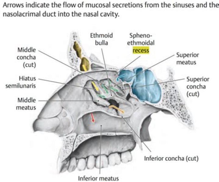
Sphenoethmoid recess is a narrow cleft bounded medially by nasal septum (perpendicular plate of ethmoid, sphenoid rostrum), laterally by the superior turbinate (ST) and superior meatus, and posterosuperiorly by the anterior face of the sphenoid body.
What is the purpose of sphenoethmoidal recess?
The sphenoethmoidal recess drains the posterior ethmoid air cells and sphenoid sinuses into the superior meatus of the nasal cavity.
Which sinus opens into Sphenoethmoidal recesses?
The sphenoid sinus ( Fig. 44.8 ) is the most posterior paranasal sinus. Its natural ostium opens into the sphenoethmoidal recess (SER). This recess lies medial and posterior to the superior turbinate, anterior to the anterior wall of the sphenoid, and medial to the nasal septum.
Where is the sphenoethmoidal recess?
The sphenoethmoidal recess lies in the angle between the ethmoid bone and the anterior surface of the sphenoid bone, above and behind the superior turbinate.What is the nasal meatus?
Identifiers. FMA. 53146. Anatomical terminology. In anatomy, the term nasal meatus can refer to any of the three meatuses (passages) through the skull’s nasal cavity: the superior meatus (meatus nasi superior), middle meatus (meatus nasi medius), and inferior meatus (meatus nasi inferior).
What are agger nasi cells?
The agger nasi cell derives its name from the Latin phrase mean- ing “nasal mound.” Arising from the most superior remnant of the first ethmoturbinal, the pneuma- tized agger nasi is a prominence or bulge along the lateral nasal wall anterosuperior to the anterior at- tachment of the middle turbinate (figure, A).
What drains in Sphenoethmoidal recess?
The sphenoethmoidal recess drains the posterior ethmoid air cells and sphenoid sinuses into the superior meatus of the nasal cavity.
What is ethmoid sinusitis?
Ethmoid sinusitis is the inflammation of a specific group of sinuses — the ethmoid sinuses — which sit between the nose and eyes. The ethmoid sinuses are hollow spaces in the bones around the nose. They have a lining of mucus to help prevent the nose from drying out.What is an ethmoid bulla?
The ethmoid bulla is the largest anterior ethmoid air cell. It is also one of the most consistent air cells in the middle meatus and is therefore a reliable anatomic landmark (Fig. 2.13). It often lacks a posterior wall, which is why it is not called a cell but rather a bony lamella with a large airspace behind it.
Where is Agger Nasi?The agger nasi (from Latin: agger meaning “mound or heap”) is a small ridge on the lateral side of the nasal cavity. It is located midway at the anterior edge of the middle nasal concha, directly above the atrium of the middle meatus.
Article first time published onWhat are paranasal air sinuses?
Paranasal sinuses are a group of four paired air-filled spaces that surround the nasal cavity. The maxillary sinuses are located under the eyes; the frontal sinuses are above the eyes; the ethmoidal sinuses are between the eyes and the sphenoidal sinuses are behind the eyes.
How do you unclog a maxillary sinus?
- Place each of your index and middle fingers on either side of your nose, just between your cheekbones and upper jaw. Try using your thumbs instead of your index fingers for stronger pressure.
- Gently massage this area using a circular motion.
- Repeat for around 30 seconds to a minute.
How do you unclog a sphenoid sinus?
- Place your index fingers on the bridge of your nose.
- Find the area between your nasal bone and the corner of the eyes.
- Hold a firm pressure in that spot with your fingers for about 15 seconds.
- Then, using your index fingers, stroke downward along the side of the bridge of your nose.
What is the name of the groove located on the back surface of the nasal bone?
The nasal bones form the roof, while the frontal process of the maxilla and lacrimal bone forms the sides of the nose. Where the maxilla and lacrimal bones connect, the lacrimal groove is formed. This groove is where the lacrimal sac resides.
What is the function of nasal chamber?
The nasal cavity lies above the bone that forms the roof of the mouth and curves down at the back to join the throat. It is divided into two sections called nasal passages. Air moves through these passages during breathing. The nasal passages filter and warm the air, and make it moist before it goes into the lungs.
What is the function of nasal septum?
The nasal septum (Latin: septum nasi) separates the left and right airways of the nasal cavity, dividing the two nostrils.
What are Haller cells?
Haller’s cells are defined as air cells situated beneath the ethmoid bulla along the roof of the maxillary sinus and the most inferior portion of the lamina papyracea, including air cells located within the ethmoid infundibulum.
What sinuses drain into middle meatus?
The middle meatus drains the frontal, anterior ethmoid, and maxillary sinuses.
What causes mucosal thickening in maxillary sinus?
Mucosal thickening is an inflammatory reaction with hyperplasia of the mucous lining of the maxillary sinus. This condition may result from harmful actions caused by trauma, infections, chemical agents, foreign body reaction, neoplasm, or airway conditions such as allergies, rhinitis, or asthma.
What is the frontal recess?
The frontal recess is bounded anteriorly by the agger nasi, laterally by the orbit, and medially by the middle turbinate. The posterior limit of the frontal recess is more variable depending upon the relationship of the ethmoid bulla or bulla lamella to the skull base.
What is Ostiomeatal complex?
The ostiomeatal complex (OMC) is the collection of structures that aids in mucus drainage and airflow between the maxillary sinus, the anterior ethmoid air cells, and the frontal sinus. It is located on the lateral wall of the nasal cavity and has several well defined borders.
What is frontal sinusotomy?
Endoscopic frontal sinusotomy involves the use of telescopes and cameras through the nose to reach the anterior ethmoid sinus, and then the frontal sinus above. These areas are opened and disease is removed.
What drains into ethmoid bulla?
Hiatus semilunaris receives drainage from the ethmoid bulla. The maxillary sinus and often the frontal sinus, depending on the superior attachment of the uncinate process, drain into the ethmoid infundibulum.
What is ethmoid infundibulum?
The ethmoid infundibulum is a curved cleft of the ethmoid bone which leads into the anterior portion of the hiatus semilunaris. It is bordered medially by the uncinate process and laterally by the orbital plate of the ethmoid.
What is a ethmoid bone?
The ethmoid bone is an unpaired cranial bone that is a significant component of the upper nasal cavity and the nasal septum. The ethmoid bone also constitutes the medial orbit wall.
What causes ethmoid inflammation?
Ethmoid sinusitis is often caused by allergies or upper respiratory infections like colds or flu. Nasal congestion from common bacteria and viral infections can restrict air into the ethmoid sinuses. Other possible causes can include larger nasal polyps or any condition that restricts the airflow in the nasal passages.
What does ethmoid sinus feel like?
Symptoms of ethmoid sinusitis You may have pain between the eyes and tenderness when touching the bridge of your nose. Other symptoms of sinusitis include: facial swelling. runny nose lasting longer than 10 days.
Can ethmoid sinuses cause headaches?
Sinus headache typically occurs in the area of the sinuses (see Figure 1)—in the area of the cheeks (maxillary sinus), bridge of the nose (ethmoid sinus), or above the eyes (frontal sinus). Less often it may refer pain to the top or back of the head (sphenoid sinus—see Figure 2 ).
What is atrium of middle meatus?
The middle meatus is below and lateral to the middle concha, and is continued anteriorly into a shallow depression, situated above the vestibule and named the atrium of the middle meatus. On raising or removing the middle concha the lateral wall of this meatus is fully displayed.
What is right concha bullosa?
Concha bullosa happens when one of the conchae, or turbinates, inside your nose becomes filled with a pocket of air. This is also known as pneumatization of the turbinate. There are three pairs of conchae in your nose on either side of the septum. Your septum is the structure that divides your nose in half.
What is paradoxical middle turbinate?
Paradoxical middle turbinate is a rare developmental cause of nasal obstruction. It refers to an inferomedially curved middle turbinate edge with the concave surface facing the nasal septum and usually occurs bilaterally.